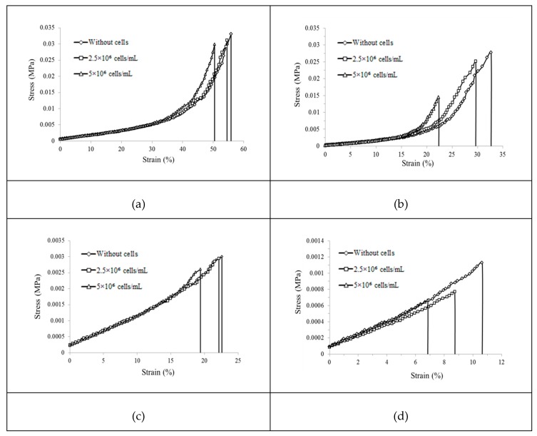
Figure 3.
Stress–strain curves of the GelMA hydrogels with different GelMA concentrations at different incubation times: (a) 10% GelMA at 0 h, (b) 10% GelMA at 96 h, (c) 5% GelMA at 0 h, and (d) 5% GelMA at 96 h (a diamond represents without cells, a square represents 2.5 × 10⁶ cells/mL, and a triangle represents 5 × 10⁶ cells/mL).

