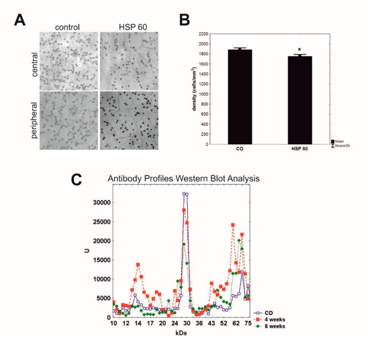
Figure 3.
(A) Retinal ganglion cells were labeled by Brn3a in retinal flatmounts of rats immunized with HSP60 and controls. The central and peripheral part of retina (40x magnification) were analyzed. (B) After eight weeks, rats immunized with HSP60 showed a significant loss of retinal ganglion cells in comparison to control animals (CO; *p = 0.02). Values are mean ± SEM. (C) Mean antigen-antibody reactivity of the serum of the HSP60 group at four and eight weeks and control group (CO) was plotted against the corresponding molecular weight of the retinal antigen. Complex antibody regulations could be detected four and eight weeks after immunization with HSP60. These changes altered according to the time after immunization. [50].

