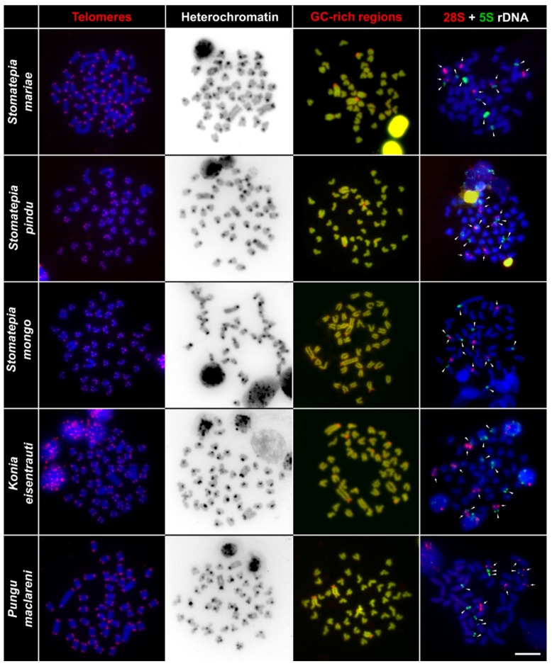
Figure 5.
Comparative chromosome analyses of Stomatepia mariae, St. pindu, St. mongo, Konia eisentrauti, and Pungu maclareni. First column: DAPI-stained chromosomes (blue) with telomere repeat hybridization signals (red) located only in the telomeric region assuming no chromosome rearrangements. Second column: inverted DAPI-stained C-banding pattern highlights clusters with constitutive heterochromatin located in the pericentromeric regions. Third column: DAPI-stained metaphase chromosomes (green), signals of GC-rich sites (red). Fourth column: DAPI stained metaphase chromosomes (blue), with six 28S rDNA (red, highlighted by arrows), and eight 5S rDNA (green, highlighted by arrowheads) hybridization signals in each species. Bar equals 10 µm.

