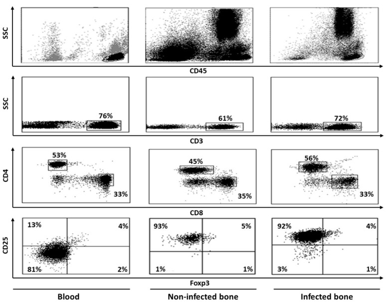
Figure 4.
Flow cytometric dot plot images showing staining patterns of T cell populations in human blood, non-infected bone and infected bone. Peripheral blood mononuclear cells (PBMCs) were separated from blood by ficoll and bone cells were isolated by cutting bone samples into fine pieces, vortexing, filtering, and centrifugation. Cells were then labeled with various monoclonal antibodies, both extracellular as well as intracellular, and analyzed by flow cytometer and doing sequential analysis. Cells were first gated on CD45 and then CD3+ population were gated on these CD45+ cells. Finally, CD4+ and CD8+ T cell populations were identified by gating on CD3+ cells. Expressions of other markers were studied on these CD4 and CD8 T cell populations. CD4+ cells that were positive for CD25 and Foxp3 double markers were considered to be Tregs. Bone tissues that were without any infection were considered non-infected, whereas those with reported bacterial infection were considered to be infected.

