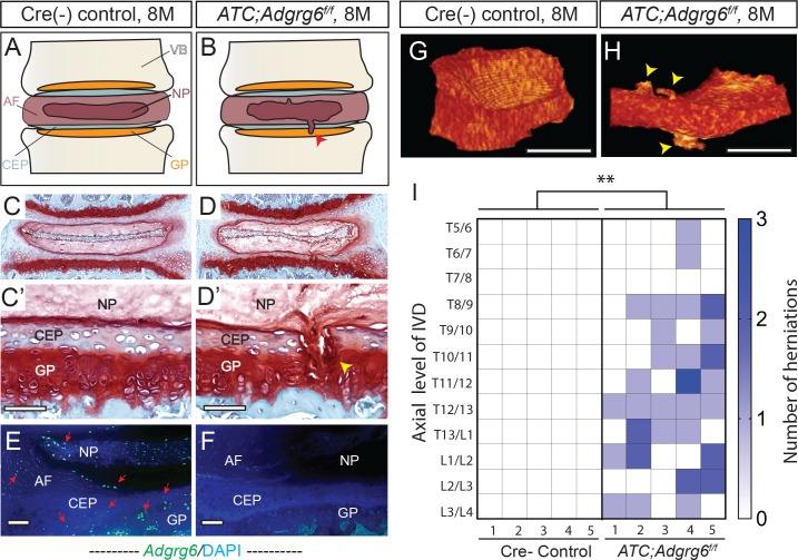
Fig 1. Adult ATC;Adgrg6f/f mutant mice display endplate-oriented herniations of the IVD.
(A and B) Schematic of endplate-oriented herniations (B, red arrowhead) observed in ATC;Adgrg6f/f mutant mice (B), in contrast to a typical wild-type IVD (A) at 8 months of age. (C-D’) Representative midline-sectioned 8-month-old mouse IVDs stained with Safranin-O/Fast green (SO/FG) (induced from P1-P20, n = 4 for each group). (E, F) Adgrg6 riboprobe FISH (green fluorescence) at 8 months (induced from P1-P20, n = 3 for each group). (G, H) Representative reconstructions of contrast-enhanced μCT of Cre (-) control (G) and ATC;Adgrg6f/f mutant (H) IVDs at 8 months of age (induced from E0.5-P20, n = 5 for each group). Endplate-oriented herniations are observed in SO/FG stained sections (D', yellow arrowhead) and by contrast-enhanced μCT (H, yellow arrowheads). (I) Heat map of contrast-enhanced μCT data from five Cre (-) control and five ATC;Adgrg6f/f mutant mouse spines, plotting the axial level of the IVD (left axis) and the number of herniations (right axis) observed in each mouse. (**p≤0.01, two-tailed Student's t Test.) Scale bars: 50μm in (C’, D’), 100μm in (E, F), and 500μm in (G, H). AF- annulus fibrosis, CEP- cartilaginous endplate, GP- growth plate, NP- nucleus pulposus, and VB- vertebral body.

