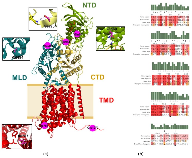
Figure 1.
Sequence and structure information on variant NPC1 protein. (a) The structure of NPC1 (PDB: 3JD8) indicated the location of the studied residues on NPC1 structure. Functional domains of the protein are indicated in different colors. NTD, N-terminal domain; MLD, middle luminal domain; CTD, C-terminal domain; and TMD, transmembrane domain. The cholesterol molecule is shown as an orange sphere. Intra and interdomain contacts of the mutation residues are shown in the insets. Hydrogen bonds are indicated as a blue dashed line. Protein structural analysis was performed with PyMol software (Schrödinger LLC, Mannheim, Germany). (b) Protein sequence alignment of the studied NPC1 residues from human with the NPC1 from mouse (Mus muculus), zebrafish (Danio rerio), and fruit fly (Drosophila melanogaster), the residues are shaded based on their levels of conservation in the alignment. The sequences were aligned using multiple sequence viewer (Schrödinger LLC, Mannheim, Germany).

