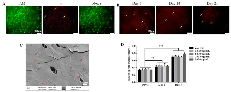
Figure 2.
Effect of TGF-β3/CS on the growth and proliferation of primary human periodontal ligament stem cells (hPDLSCs). (A) Calcein-AM/ propidium iodide (PI) double staining of hPDLSCs on TGF-β3/CS after 3 d of culture in vitro. Live cells (red arrow) are stained by AM (green), and dead cells (yellow arrow) are stained by PI (red) (×100). Scale bar represents 100 μm. (B) TGF-β3/CS with hPDLSCs (white arrow) implanted in Sprague Dawley rats for 7, 14, and 21 d and then stained with CM-Dil (red). Cell survival was observed under a fluorescence microscope (×100). Scale bar represents 100 μm. (C) SEM photomicrographs of hPDLSCs (red arrow) in CS for 7 d (×200). Scale bar represents 40 μm. (D) hPDLSC growth in TGF-β3/CS was measured by CCK-8 assays (mean ± SD; n = 5). ** p < 0.01 vs. control; *** p < 0.001 vs. control.

