Figure 1.

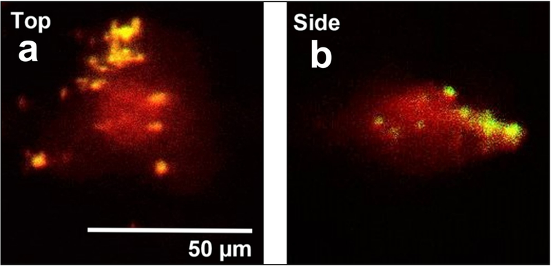
Representative images showing CWP (yellow) attachment to surface of healthy human FLS (34-M) (red) after 6 days in static culture. Top and side views acquired via quasi-3D microscopy.

Representative images showing CWP (yellow) attachment to surface of healthy human FLS (34-M) (red) after 6 days in static culture. Top and side views acquired via quasi-3D microscopy.