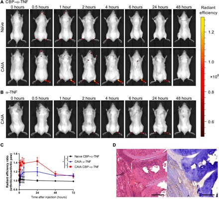
Fig. 2. CBP–α-TNF accumulated in the inflamed paw.

Arthritis (CAIA) was induced selectively in the right hind paw by passive immunization of anticollagen antibodies, followed by subcutaneous injection of LPS at the right hind footpad and phosphate-buffered saline at the left hind footpad. On the day following LPS injection, Cy7-labeled CBP–α-TNF and Cy7-labeled α-TNF were intravenously injected into naïve and CAIA mice. Representative images of accumulation in arthritic or nonarthritic paws of mice injected with CBP–α-TNF (A) and α-TNF (B) are shown. (C) Changes in radiant efficiency ratio of the arthritic paw (right hind) to the nonarthritic paw (left hind) in naïve and CAIA mice (n = 3 to 4, means ± SD). *P < 0.05 compared with the area under the radiant efficiency ratio-time curve from 0 to 72 hours of each treatment group (Tukey’s multiple comparison test). (D) Representative histology images of joints in CBP–α-TNF–injected CAIA mouse [left, hematoxylin and eosin (H&E) staining; right, immunohistochemistry staining against anti-rat immunoglobulin G (IgG)]. Scale bars, 200 μm.
