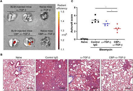
Fig. 4. Localization in lung and efficacy of CBP–α–TGF-β in the BLM-induced pulmonary fibrosis model.

Mice were intranasally instilled with 50 μg of BLM sulfate on day 0. (A) Cy7-labeled CBP–α–TGF-β or Cy7-labeled α–TGF-β was intravenously injected into naïve mice or BLM-injected mice on day 7. The lung was harvested 4 hours after the fluorescence injection, and fluorescence intensity was measured. (B and C) Control IgG, unmodified α–TGF-β, or CBP–α–TGF-β at a dose of 50 μg per mouse three times a week from day 7. On day 21, the left lung lobe was fixed and provided to histological analysis. (B) Representative image of Masson’s trichrome staining of the lung on day 21 in each treatment group. Scale bars, 200 μm (C) Lung fibrosis was scored 0 to 8 by Ashcroft scoring as described in Materials and Methods. *P < 0.05 compared with the scores of each treatment group (Tukey’s multiple comparison test).
