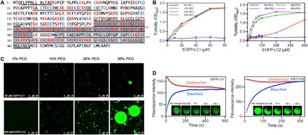
Fig. 1. Sequences and LLPS of LC1 and LC2 of U1-70K.

(A) U1-70K amino acid sequence with basic residues in red and acidic residues in blue. Disordered sequences by predictions were underlined, and LC domains were highlighted in gray. (B) Turbidity demonstrated phase separation of LC1 and LC2. (C) Confocal microscopy showed droplet formation of 60 μM LC1 and 200 μM LC2. (D) FRAP of the droplets formed by LC1 and LC2.
