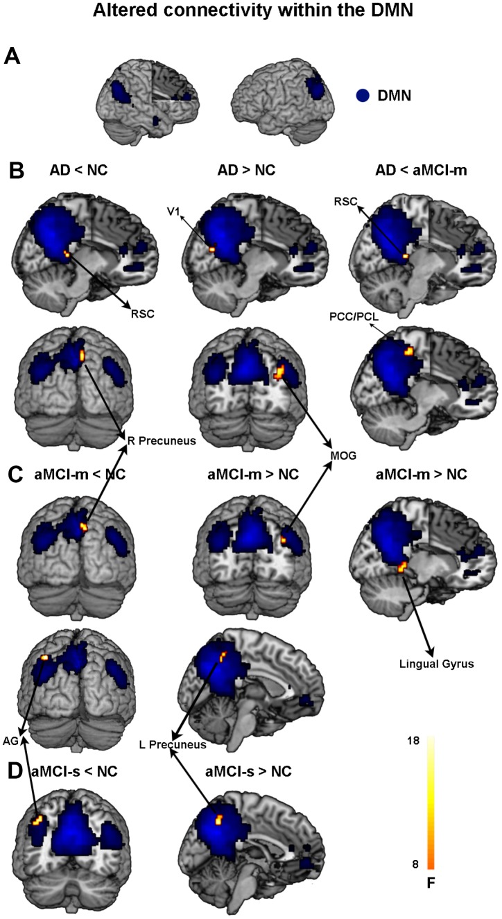
Figure 1.
Altered functional connectivity within the DMN among the four groups. (A) Spatial distribution of the DMN (FWE correction). (B) Regions showing hypo- and hyperconnectivity within the DMN in the AD group; (C) Regions showing hypo- and hyperconnectivity within the DMN in the aMCI-m group; (D) Regions showing hypo- and hyperconnectivity within the DMN in the aMCI-s group. Abbreviations: DMN: default mode network; RSC: retrosplenial cortex; V1: primary visual cortex; MOG: middle occipital gyrus; PCC: posterior cingulate cortex; PCL: paracentral lobule; AG: angular gyrus; AD: Alzheimer's disease; aMCI-s: single-domain of amnestic mild cognitive impairment; aMCI-m: multiple-domain of amnestic mild cognitive impairment; NC: normal controls.

