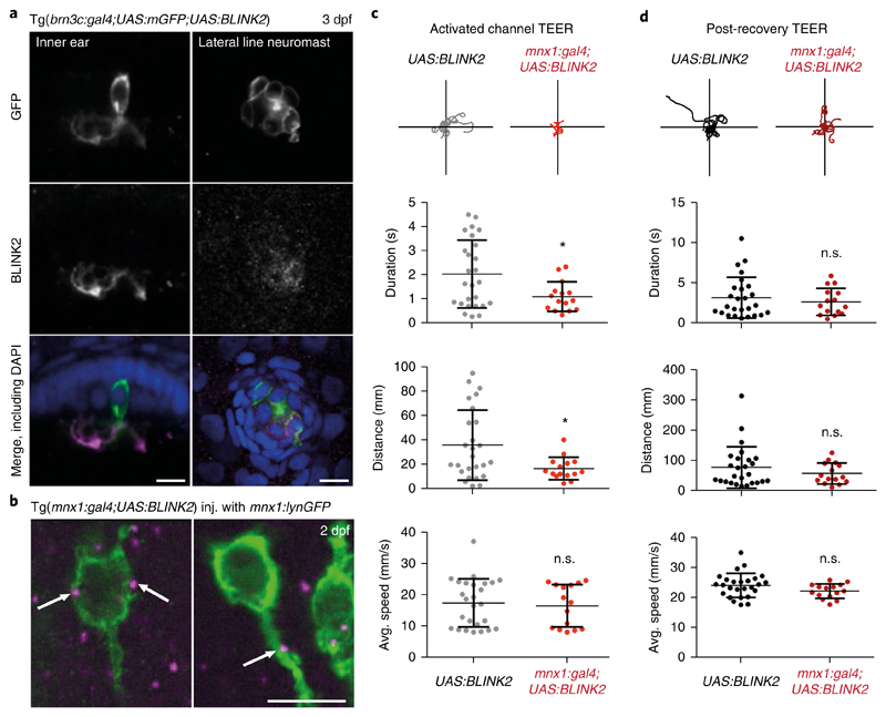
Fig. 4. BLINK2 expression and functional silencing in zebrafish.
a, Left, immunohistochemistry in 3-dpf (days post-fertilization) embryos, showing a hair cell of the inner ear labeled for membrane-targeted GFP (green in the merged image) and the BLINK2 channel (magenta in the merged image), both expressed under the control of brn3c:gal4. Right, neuromast cells from the same Tg(brn3c:gal4;UAS:mGFP) line labeled in the same way. Embryos were counterstained with DAPI (blue). Scale bars, 10 μm. Similar results were obtained in 3 independent experiments. b, Immunohistochemistry on whole 2-dpf embryos showing cell bodies and part of the axons of primary motor neurons stained by membrane-targeted GFP (green) and BLINK2 (magenta). White arrows indicate BLINK2 immunoreactivity at the plasma membrane and axonal tract. Genotypes are as indicated. GFP was expressed in subsets of motor neurons only. Scale bars, 10 μm. Similar results were obtained in 3 independent experiments. c, Touch-evoked escape response assay (TEER) in Tg(mnx1:gal4;UAS:BLINK2) and Tg(UAS:BLINK2) embryos. Embryos were assayed after a 20-min activation of the channel with blue light. Swim duration, distance and average speed were 2.03 ± 0.28 s, 35.80 ± 5.65 mm and 17.49 ± 1.50 mm/s, respectively, in control animals and 1.10 ± 0.16 s, 16.62 ± 2.38 mm and 16.60 ± 1.75 mm/s in BLINK2-expressing animals. Traces for 10 escape episodes are shown for each condition. n = 26 larvae for Tg(UAS:BLINK2) and n = 15 larvae for Tg(mnx1:gal4;UAS:BLINK2). Data are presented as the average (center line) ± s.d.; P values are, respectively, 0.019, 0.017 and 0.071. d, TEER assay in the same animals as in c after 1 h of rest in the dark. Swim duration, distance and average speed were 3.18 ± 0.50 s, 77.12 ± 13.42 mm and 24.03 ± 0.79 mm/s, respectively, in control animals and 2.67 ± 0.44 s, 57.03 ± 8.90 mm and 22.08 ± 0.63 mm/s in BLINK2-expressing animals. P values are, respectively, 0.48, 0.29 and 0.099. *P ≤ 0.05 (two-sided t-test). n.s., not significant.

