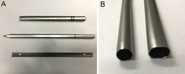
Figure 2.
A: The images in order from top to bottom are of an oval dilator, oval sleeve, and a J-shaped nerve retractor (Robert Reid Inc., Tokyo, Japan). B: The left image is of the oval sleeve for percutaneous endoscopic discectomy, and the right image is of the elliptical sleeve for percutaneous endoscopic transforaminal lumbar interbody fusion. The short axis was set as the same size.

