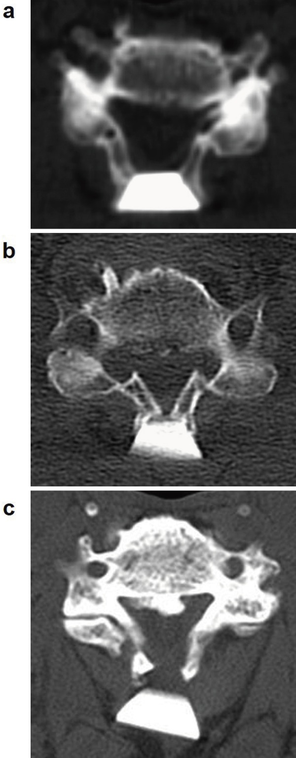
Figure 3.

Computed tomograms of the “open-door” lamina after midline-splitting laminoplasty. (a) Ideally opened lamina with a rhomboid-shaped hydroxyapatite spacer. (b) Fractured “open-door” lamina sinking toward spinal canal. (c) Displaced hydroxyapatite spacer causing closure of the opened lamina.
