Figure 1.
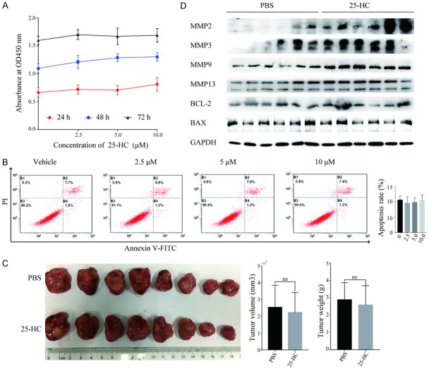
25-HC has no effects on HepG2 cells proliferation and apoptosis both in vitro and in vivo. (A) HepG2 cells were treated with the indicated concentrations of 25-HC for 24, 48 or 72 hours and CCK-8 assay was performed. Results are shown as the cells absorbance at 450 nm. (B) HepG2 cells were treated with the indicated concentrations of 25-HC for 48 hours followed by the flow cytometry analysis of cell apoptosis. The percentage of FITC positive cells indicating apoptotic HepG2 cells were calculated and statistically analyzed. (C) Nude mice were inoculated subcutaneously with HepG2 cells (2 × 10⁶) followed by PBS or 25-HC treatment intraperitoneally. The volume of the tumor and the body weight was monitored. Tumors were photographed at the end of the experiment. (D) Proteins of xenograft tumors of two groups were extracted and the expressions of MMP2, MMP3, MMP9, MMP13, Bcl-2 and Bax were determined by Western blotting. Results were obtained from 3 independent experiments (A and B) or 2 independent experiments (C and D) and are expressed as the means ± SEM. Statistical significance in (C) was determined by Student’s t-test. ns, not significant.

