Figure 2.
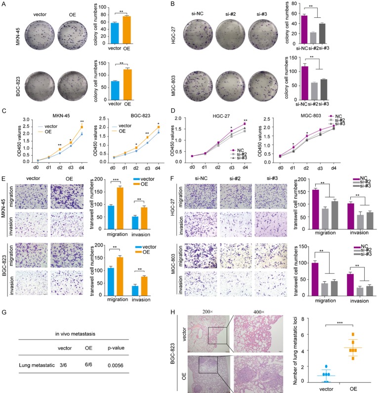
(A, B) Colony formation assays with representative images of MKN-45 and BGC-823 cells infected with the lentivirus expressing onclncRNA-626, as well as HGC-27 and MGC-803 cells transfected with two independent onclncRNA-626 siRNAs. (C, D) CCK-8 assays of MKN-45 and BGC-823 cells infected with the lentivirus expressing onclncRNA-626 and HGC-27 and MGC-803 cells transfected with two independent onclncRNA-626 siRNAs. (E, F) Transwell migration and invasion assays of MKN-45 and BGC-823 cells as well as in HGC-27 and MGC-803 cells after manipulating onclncRNA-626 expression. (G) Hematoxylin-eosin staining of lung sample sections with metastatic nodules obtained from nude mice after injection with BGC-823 cells infected with the lentivirus expressing onclncRNA-626. (H) Statistics analysis of the metastatic foci in the lung detected by hematoxylin-eosin staining. Data are shown as the mean ± SEM, n=3 in (A, B, E and F), n=6 in (H). *P<0.05, **P<0.01, ***P<0.001. Bars: 100 μM.

