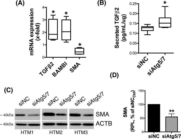
Figure 2.
Decreased expression of fibrotic markers in siAtg5/7-transfected TM cells. (A) qPCR analysis confirming changes in expression levels of TGFβ2, BAMBI and SMA genes in siAtg5/7-transfected TM cells. (B) Levels of total TGFβ2 (latent and active) in culture media from siAtg5/7-transfected TM cells quantified by ELISA and normalized with total μg of corresponding whole cell lysates. (C) Protein levels of SMA as evaluated by western blot in whole-cell lysates (5 μg) from three independent human TM primary cells transfected for 72 h with siAtg5/7. (D) Mean SMA relative protein levels quantification as calculated from densitometric analysis of the bands, expressed as percentage of siNC controls. β-actin was used as loading control. *p < 0.05, **p < 0.01, t-test, n = 3. RPL: Relative protein levels.

