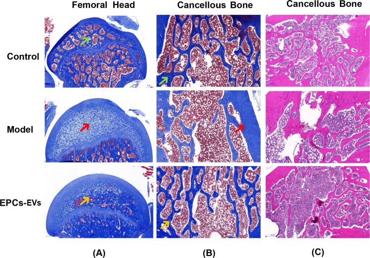
Figure 4.
Histopathological staining of femurs in mice with SIOP following EPC-EV treatment. After the treatment of each group, all animals were sacrificed, and their bilateral femurs were recovered, fixed, and decalcified, histopathological sections were prepared for staining. (A) Representative pictures of Masson staining in the coronal plane of the femoral head from each group. The green arrow shows the normal structure of the femoral head. The red arrow shows the necrotic tissue that replaced the normal bone structure and marrow. The yellow arrow shows the partially reduced necrosis and recovered blood supply. (B) Representative pictures of Masson staining in the cancellous bone of femurs from each group. The green arrow shows normal collagen staining following Masson staining. The red arrow shows that there was less abundant collagen with a light blue colour after Masson staining. The yellow arrow shows the partial recovery of collagen abundance. (C) Representative pictures of HE staining in a coronal plane of the cancellous bone from each group.

