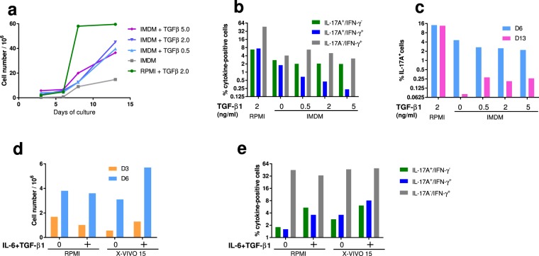
Figure 4.
Choice of culture medium for the expansion of Th17 cells. (a) Cell growth after 3, 6, 8 or 13 days in RPMI (+FCS and 2 ng/mL TGF-β1) or IMDM (+10% KnockOut Serum Replacement) with different concentrations of recombinant human TGF-β1. (b) Proportions of IL-17A, IFN-γ and double positive cells after 6 days of culture as a function of TGF-β1 concentration (0 to 5 ng/mL). Results are means from the cells of two cows. (c) Proportions of IL-17A+ cells after 6 and 13 days of culture as a function of TGF-β1 concentration (0 to 5 ng/mL). Results are means from the cells of two cows. (d) Comparison of cell growth in RPMI and X-VIVO™ 15 media with or without polarizing cytokines (40 ng/mL IL-6 and 2 ng/mL TGF-β1), after 3 and 6 days of culture. (e) Proportions of cells IL-17A+ and IFN-γ+ (ICS) with or without polarizing cytokines (40 ng/mL IL-6 and 2 ng/mL TGF-β1) after 6 days of culture. Results are means from two cows.

