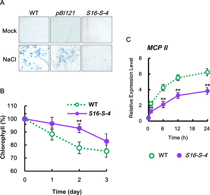
Figure 2.
Determination of cell damage in transgenic tobacco leaves under salt stress. (A) Mature tobacco leaves were treated with 200 mM NaCl for the indicated time points in WT, transgenic plants with vector control (pBI121), and S16-S-4; necrotic areas were then stained with trypan blue and imaged with a digital camera. (B) Changes in chlorophyll contents in leaves of WT and transgenic plants after salt stress. (C) Transcription levels of tobacco Metacaspase II (MCPII) gene in tobacco plants after salt stress. Real-time qPCR analysis of MCPII transcription using total RNAs from tobacco leaves. Transcription levels are expressed relative to the reference gene β-actin after real-time qPCR. Relative messenger RNA (mRNA) expression levels are expressed as means ± SD. The photographs represented (A) are from one representative experiment in five independent experiments with more than three leaves after verifying the reproducibility of the results. Data (B, C) were generated from one representative experiment with three independent biological replicates after verifying the reproducibility of the results in three experiments. An asterisk indicates a significant difference between WT and transgenic plants (**P < 0.01).

