Figure 1.
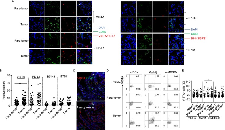
VISTA protein is mainly expressed by intratumoral myeloid cells. (A) Immunofluorescence analyses demonstrating the expression of VISTA, PD-L1, B7-H3 and B7S1 together with DAPI and CD45 in paired tumors and para-tumors. (B) Quantifications of VISTA, PD-L1, B7-H3 and B7S1 by immunofluorescence staining were shown (n = 47). **P < 0.01. (C) Immunofluorescence analyses demonstrating VISTA expression on tumor cells. (D and E) Representative figures and summarized data showing percentage of VISTA+ cells in mDCs, monocytes/macrophages, monocytic MDSCs from PBMC, para-tumors and tumors of ccRCC patients (n = 53). *P < 0.05

