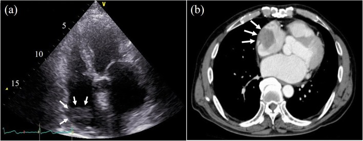
Fig. 1.
a A 45 × 27 mm mass (white arrows) was observed as an area of low density near the right atrial free wall on preoperative contrast-enhanced computed tomography. b A round floating mass (white arrows) with a relative low echogenicity was recognized in the right atrium in the preoperative four-chamber view of transthoracic echocardiography

