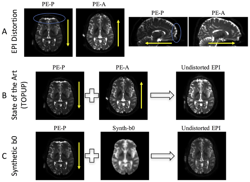
Figure 2.
Distortion and correction in diffusion MRI. (A) EPI susceptibility distortion occurs along the phase encode direction, with phase encoding in the posterior (PE-P) direction leading to displacements of identical distance but opposite direction of that in the anterior (PE-A) direction. (B) State of the art distortion correction (topup) typically uses distortions in two opposite directions to iteratively estimate the undistorted image. (C) The proposed method uses an undistorted T1-weighted image to synthesize an undistorted volume with b0 contrast, which can be used to correct the distorted (in this case, PE-P) image without requiring an additional phase encoding acquisition. The blue circles highlight areas of observable signal distortion. Yellow arrows indicate phase encode direction.

