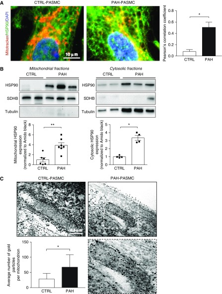
Figure 1.
HSP90 preferentially accumulates in mitochondria of pulmonary arterial hypertension–pulmonary artery smooth muscle cells (PAH-PASMCs). (A) Representative images of HSP90 (green) localization in human control and PAH-PASMCs labeled with MitoTracker Red. Quantification of HSP90-MitoTracker Red colocalization is shown in the right panel. (B) Representative Western blotting analysis and corresponding densitometric analyses of mitochondrial and cytosolic fractions obtained from control (n = 4–6) and PAH-PASMC (n = 4–7) lysates and probed for HSP90. To assess fraction purity, antibodies for succinate dehydrogenase complex, subunit B and α-tubulin were used. (C) Representative images of control PASMCs and PAH-PASMCs immunogold labeled with an anti-HSP90 antibody and corresponding quantification of the average number of gold particles per mitochondria in control PASMCs and PAH-PASMCs. The lower right image is an enlargement of the area inside the dashed box in the upper right image. Data are presented as mean ± SEM in A and B or mean ± SD in C. *P < 0.05; **P < 0.01. CTRL = control; SDHB = succinate dehydrogenase complex, subunit B.

