Figure 2.
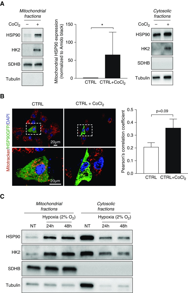
Hypoxia induces mitochondrial HSP90 accumulation in control pulmonary artery smooth muscle cells (PASMCs). (A) Representative Western blotting analysis of mitochondrial and cytosolic fractions obtained from control PASMCs (n = 4) exposed or not to 500 μM cobalt chloride (CoCl2) for 2 hours and probed for HSP90. Data are presented as mean ± SEM; *P < 0.05. (B) Representative images of control human PASMCs transfected with a plasmid encoding a green fluorescent protein (GFP)-tagged HSP90 and stimulated or not with CoCl2 (500 μM) for 2 hours. Cells were labeled with MitoTracker Red. The left and right lower images are enlargements of the area inside the dashed boxes in the left and right upper images, respectively. Quantification of GFP-MitoTracker Red colocalization is shown in the right panel. Data are presented as mean ± SEM. (C) Western blotting analysis of mitochondrial and cytosolic fractions obtained from control PASMCs exposed or not to 2% O2 for 24 or 48 hours and probed for HSP90. Immunoblotting with SDHB and with α-tubulin was used to assess purity of the mitochondrial and cytoplasmic fractions, respectively. Hexokinase 2 (recruited to mitochondria under hypoxic stimulation) was used as a positive control of hypoxia exposure or CoCl2 treatment. CTRL = control; HK2 = hexokinase 2; NT = nontreated; SDHB = succinate dehydrogenase complex, subunit B.

