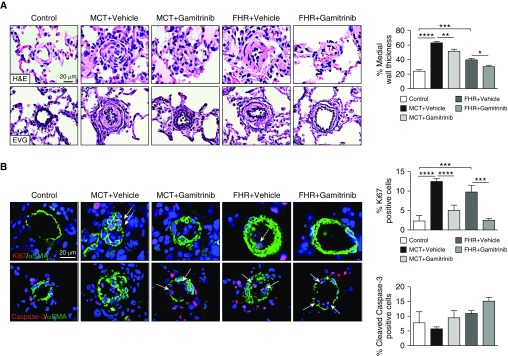
Figure 7.
Mitochondrial HSP90 inhibition using Gamitrinib improves vascular remodeling in monocrotaline (MCT)-induced PAH and fawn-hooded rats (FHRs). (A) Representative images of distal pulmonary vessels stained with hematoxylin and eosin or Elastica von Gieson and corresponding quantification of vascular remodeling. (B) Proliferation (Ki67) and apoptosis (cleaved Caspase-3) were evaluated in lungs of control, MCT + vehicle, MCT + Gamitrinib, FHR + vehicle, and FHR + Gamitrinib rats. Representative images of distal pulmonary vessels labeled with Ki67 (top) and cleaved Caspase-3 (bottom) in red. Vascular smooth muscle cells were labeled using α-smooth muscle actin staining (green). Graphs on the right represent the percentage of cells positive for Ki67 or cleaved Caspase-3 in distal pulmonary vessels. Arrows indicate positive cells. Data are presented as mean ± SEM; *P < 0.05; **P < 0.01; ***P < 0.001; ****P < 0.0001. αSMA = α-smooth muscle actin; EVG = Elastica von Gieson; H&E = hematoxylin and eosin; PAH = pulmonary arterial hypertension.

