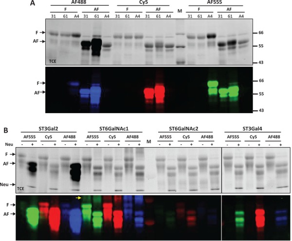
Fig. 2.

DFGL on fetal bovine fetuin and asialofetuin. All labeled samples were separated on SDS-PAGE and imaged by both TCE staining (top panels) and fluorescent imaging (lower panels). (A) Fetal bovine fetuin (F) and asialofetuin (AF) were labeled by ST3Gal1 (31), ST6Gal1 (61) and ST6GalNAc4 (A4) with Alexa-Fluor®488 (AF488), Cy5 and Alexa-Fluor®555 (AF555). (B) Labeling of fetuin and asialofetuin samples by ST3Gal2, ST6GalNAc1, ST6GalNAc2 and ST3Gal4 with AF555, Cy5 and AF488. Asialofetuin in (A) was purchased from Sigma Aldrich. Asialofetuin in (B) was generated from fetuin by addition of C.p neuraminidase (Neu) to the labeling reactions. ST6GalNAc1 exhibited self-labeling [indicated with arrow in (B)]. Same amount of protein (2.5 μg) was loaded into each lane; however, due to the presence of multiple benzene rings, Alexa Fluor® fluorophore-labeled samples exhibit significantly increased band intensities in TCE images. M represents molecular marker.
