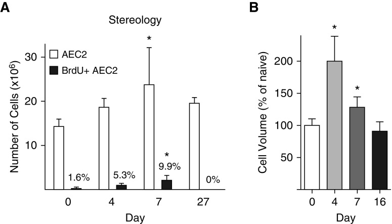
Figure 1.
Alveolar epithelial type 2 cell (AEC2) expansion and volume measured by stereology. SPCCreERT2+/−;mTmG+/− mice were treated with LPS or saline as a control. Mice were administered bromodeoxyuridine (BrdU) 24 hours before being harvested. Lungs were inflation-fixed, cut into 3-mm slabs, divided into two blocks, and processed. One paraffin block was exhaustively cut into 30-μm sections. Sections were immunostained for green fluorescent protein and BrdU and imaged at ×63. (A) Cells were counted with an optical fractionator. The total number of AEC2s is indicated by the open bars; numbers of actively cycling (BrdU+) AEC2s are indicated by the solid bars. The percentage of total AEC2s that are BrdU+ is shown above the solid bars. (B) The volume of individual cells was measured by the nucleator method and is reported as a percentage of the naive animal. There was no difference in shrinkage in the x–y plane between naive and LPS-treated lungs (data not shown). Means and SD are shown. One-way ANOVA with Bonferroni post hoc analysis was performed. *P < 0.05 compared with naive lung.

