-
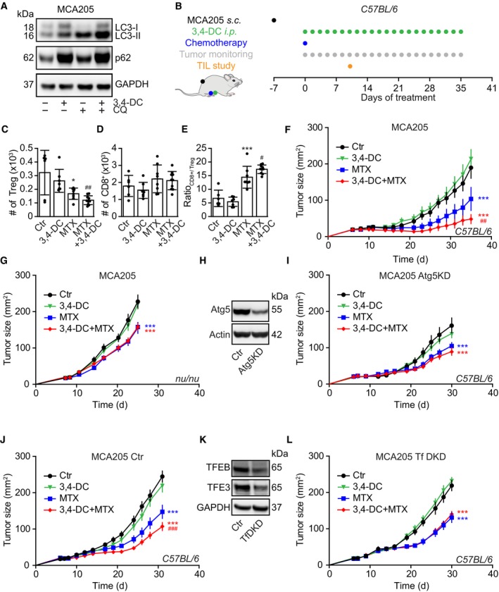
A
Induction of autophagy in murine MCA205 fibrosarcomas. Cells were treated with 3,4‐DC alone or in combination with chloroquine, and were harvested 6 h later for immunoblot detection of LC3 lipidation.
-
B
Schematic overview of the in vivo treatment of MCA205 fibrosarcomas with mitoxantrone (MTX) and 3,4‐DC, alone or in combination.
-
C–E
Cytofluorometric characterization of tumor‐infiltrating lymphocytes (TIL), in particular FOXP3+ regulatory T cells (Treg), CD8+ cytotoxic T lymphocytes, and the ratio of CD8+ T cells over Treg.
-
F–I
Growth kinetic of MCA205 fibrosarcomas that were either wild‐type (F, G) or Atg5KD (H, I) and were evolving in immunocompetent C57Bl/6 mice (F, I) or immunodeficient nu/nu mice (G), treated as indicated in (B).
-
J–L
Immunocompetent C57Bl/6 mice were subcutaneously inoculated with TFEB/TFE3 double knockdown MCA205 cells or its scramble control cells (K). When tumors became palpable, mice were treated as indicated in (B). Tumor growth curves from mice subjected to 3,4‐DC administration alone or in combination with MTX are shown (J, L).
Data information: Asterisks indicate significant effect of MTX with respect to untreated controls (mean value ± SEM, *
P < 0.05, ***
P < 0.001; Student's
t‐test), while hash symbols refer to the comparison of the effects of MTX plus 3,4‐DC to MTX alone (
#
P < 0.05,
##
P < 0.01,
###
P < 0.001; Student's
t‐test) (
n = 6–14). Samples for immunoblots were run on one gel (A, H) or several parallel gels (K), then blotted, cut into horizontal stripes, and probed separately.
Source data are available online for this figure.