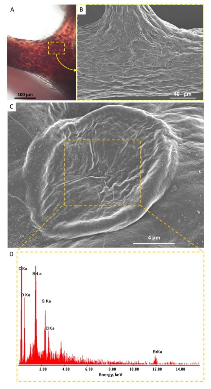
Figure 4.
Pigmental cells located within fibers of I. flabelliformis chitin are clearly visible using light microscopy (A) (see also Figure 3C). These cells are observable using SEM (B,C). Single-spot energy-dispersive X-ray spectroscopy (EDX) analysis shows strong evidence of the presence of bromine within individual pigmental cells (D).

