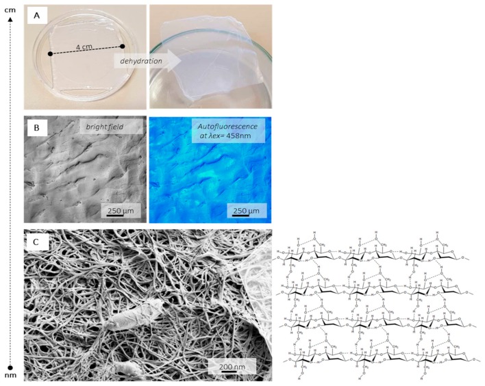
Figure 4.
Different length-scale presentation of BC: (A) photographs of wet (left) and dry (right) BC membrane, (B) confocal fluorescent microscopy (CFM) image obtained under argon laser excitation at 458 nm from bright field and fluorescence channel, utilizing the cellulose autofluorescence and (C) high magnification scanning electron microscopy (SEM) image presenting entrapped K. xylinus bacteria and cellulose backbone insert.

