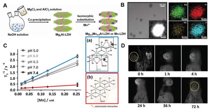
Figure 5.
(A) Schematic illustration of synthetic procedure, (B) TEM image, STEM image and the corresponding element mapping of Mn-LDH nanoparticles, (C) Plot of T-1 versus Mn concentration of Mn-LDH nanoparticles after co-incubation with different pH buffer solution at 37 °C for 4 h and 2D atomic structure models of Mn-LDH dispersed in pH 5.0 (a) and pH 7.4 (b) buffer, (D) in vivo MR imaging in the melanoma tumor-bearing mouse after intravenous injection of BSA/Mn-LDH nanomaterial within 72 h, from [26], with permission from WILEY-VCH Verlag GmbH & Co. KGaA, 2017.

