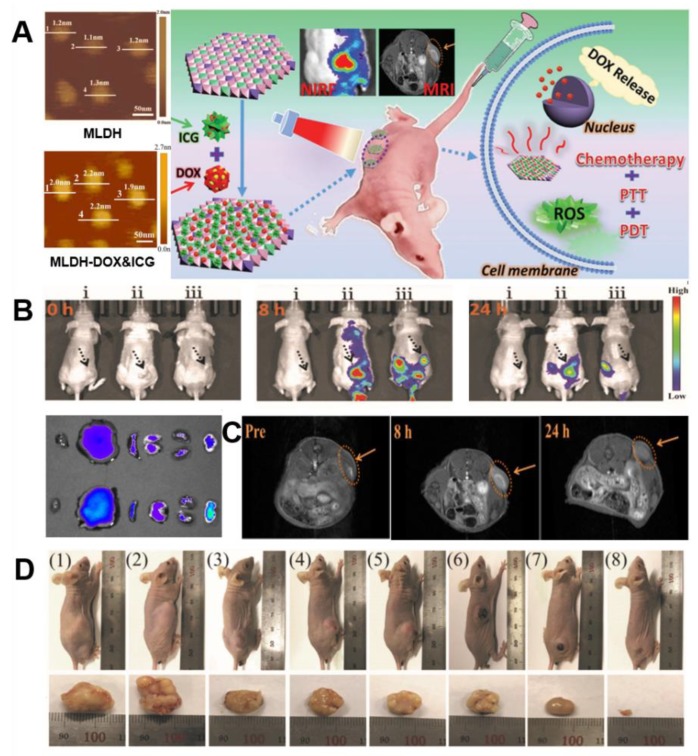
Figure 10.
(A) AFM images of Gd-MLDH and Gd-MLDH-DOX&ICG nanosheets (left), schematic illustration for MLDH-based versatile platform that can be utilized for dual modal imaging including NIRF and MR imaging with the synergistic chemo-/PT/PD combination therapy (right). (B) In vivo fluorescence imaging of nude mice bearing HepG2 tumors at different time points after i.v. injection of i: saline, ii: MLDH-DOX&ICG, and iii: DOX&ICG (tumors are pointed out by the black arrows) and NIRF images of tumor and different organs after i.v. injection of DOX&ICG (top) and MLDH-DOX&ICG (bottom) at 24 h (respectively heart, liver, spleen, lung, kidney, tumor, from left to right). (C) In vivo T1-weighted MR images at different time points after i.v. injection of MLDH-DOX&ICG (tumor locations are indicated by the orange arrows). (D) Digital photographs of the mice on day 14 after various treatments and corresponding excised tumors (respectively from (1) to (8): saline, MLDH nanosheets, DOX&ICG, MLDH-DOX&ICG, MLDH-DOX with irradiation, DOX&ICG with irradiation, MLDH-ICG with irradiation, and MLDH-DOX&ICG with irradiation), reproduced from [57], with permission from WILEY-VCH Verlag GmbH & Co. KGaA, 2018.

