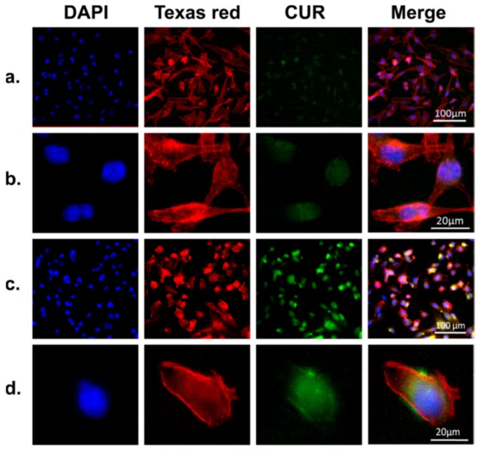
Figure 5.
Representative microscopic images of MDA-MB-231 cells incubated with free curcumin ((a) and (b), curcumin amount (10 μg/mL), equivalent to the curcumin amount in CMSPs (curcumin loaded magnetic SF core−shell nanoparticles) and CMSPs ((c) and (d), 30 μg/mL) for 4 h. The cell nucleus and cytoskeleton were stained with DAPI (blue) and Texas red (red); all images were taken with an AF6000 microscope (Leica). Comparing the images in (a) and (b) to (c) and (d), it can be seen that CMSPs significantly improve the cellular uptake of the curcumin. Reprinted from Reference [43] with permission from the American Chemical Society [2017].

