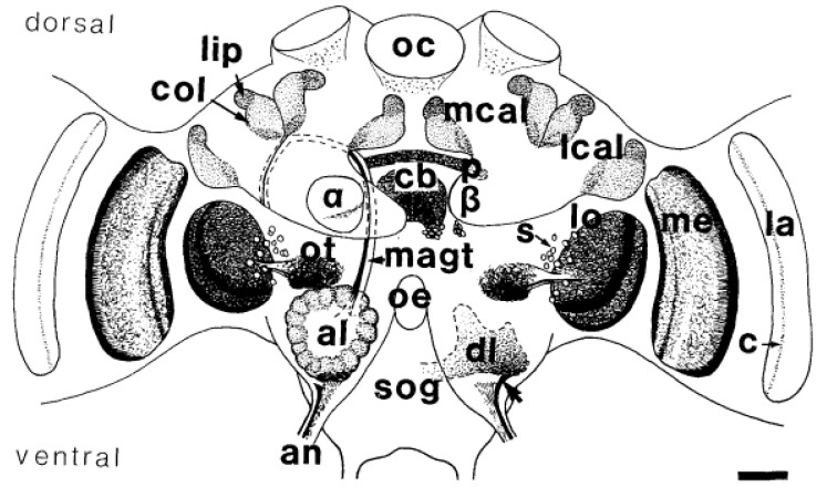
Figure 1.
Schematic drawings of acetylcholinesterase (AchE) labelling in the neuropiles and some prominent groups of somata in the bee’s brain in a frontal view. The optic lobes lamina (la), medulla (me), and lobula (lo) display layered AChE staining. Weak AChE staining was found in the synaptic plexus of the ocelli (oc). The lobula is connected to the optic tubercle (ot). AChE-positive sensory fibres (arrow) of the antennal nerve project into the dorsal lobe (dl) and the suboesophageal ganglion (sog) below the oesophagus (oe). The median antennoglomerular tract (magt) connects the glomeruli of the antennal lobe (al) with the lip area (lip) of the median (mcal) and lateral calyx (Ical) of the mushroom bodies (MB). The collar (col) is a neuropilar compartment of the calyx receiving visual input. AChE-positive fibres leave the β-lobe (β) of the mushroom body. The central complex shows AChE activity in the pons (p), central body (cb) and a group of somata (s) ventrally to the central body. C-layer (c), α-lobe (α), antennal nerve (an). Scale = 100 µm (reproduced from [31] with permission by John Wiley and Sons).

