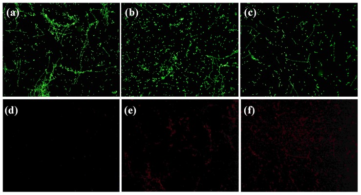
Figure 8.
Fluorescent microscope images of Candida albicans biofilms, showing (a–c) live cell distributions and (d–f) dead cell distributions. (a,d) Control biofilms, (b,e) biofilms treated with ZnFe2O4@AgNWs hybrid nanostructures for 12 h, (c,f) biofilms treated with ZnFe2O4@AgNWs hybrid nanostructures for 24 h. Measurements were made using a 10× objective lens.

