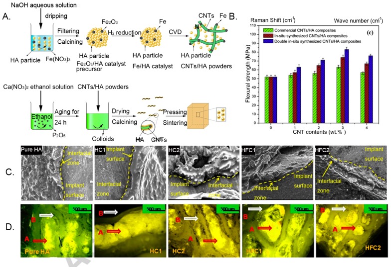
Figure 2.
Optimized bioavailability of a carbon nanotube–hydroxyapatite (CNT-HA) composite in bone engineering. (A) Schematic diagram of the preparation process of the CNT–HA composite. (B) Flexural strength of HA composites with different contents of CNTs. (C) SEM micrographs of the host–implant interface 120 days after implantation. (HA: hydroxyapatite; HC1:HA + 1% WCNT; HC2:HAC + 2% MWCNT; HFC1:HA + 1% functionalized-MWCNT, and HFC2:HA + 2% functionalized-MWCNT). (D) Fluorochrome labeling images at 120 days after implantation showing new bone (golden yellow) and old bone (deep sea green). Reproduced with permission from [147,148]. Elsevier, 2016.

