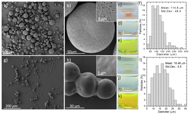
Figure 1.
SEM photographs showing the morphology of empty microparticles based on: (a,b) Eudragit RS 100; (g,h) Eudraguard biotic; (c) Optical imaging of an Eudragit-based microparticle suspension in water; (d) Optical imaging of the supernatant collected from a Eudragit-based microparticle suspension after 2 h at 37 °C under simulated gastric conditions; (e) Optical imaging of the supernatant collected from a Eudragit-based microparticle suspension after 6 h at 37 °C under simulated intestinal conditions; (f) Size distribution of Eudragit RS 100 microparticles obtained (N = 150); (i) Optical imaging of a Eudraguard-based microparticle suspension in water; (j) Optical imaging of the supernatant collected from a Eudraguard-based microparticle suspension after 2 h at 37 °C under simulated gastric conditions; (k) Optical imaging of the supernatant collected from a Eudraguard-based microparticle suspension after 6 h at 37 °C under simulated intestinal conditions; (l) Size distribution of Eudraguard biotic microparticles obtained (N = 150). Std. Dev. = Standard Deviation.

