Figure 5.
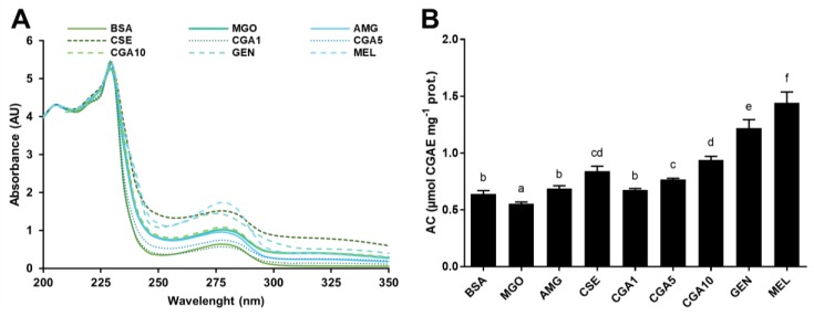
UV-Vis spectra (A) and antioxidant capacity (B) of the high molecular weight protein fractions (>30 kDa) isolated from the samples of the BSA-MGO glycation system (72 h agitation, 37 °C, pH 7.4) BSA: negative control; MGO: BSA + methylglyoxal; AMG: BSA + methylglyoxal + aminoguanidine; CSE: BSA + methylglyoxal + CSE (12,5 mg mL−1); CGA1: BSA + methylglyoxal + 1 mmol L−1 CGA; CGA5: BSA + methylglyoxal + 5 mmol L−1 CGA; CGA10: BSA + methylglyoxal + 10 mmol L−1 CGA; GEN: BSA + methylglyoxal + 5 mmol L−1 genistein; MEL: BSA + methylglyoxal + 5 mmol L−1 melatonin. Results are presented as mean ± SD (n = 3). Bars with different letters denote significant differences (p < 0.05) when subjected to Tukey multiple range test.

