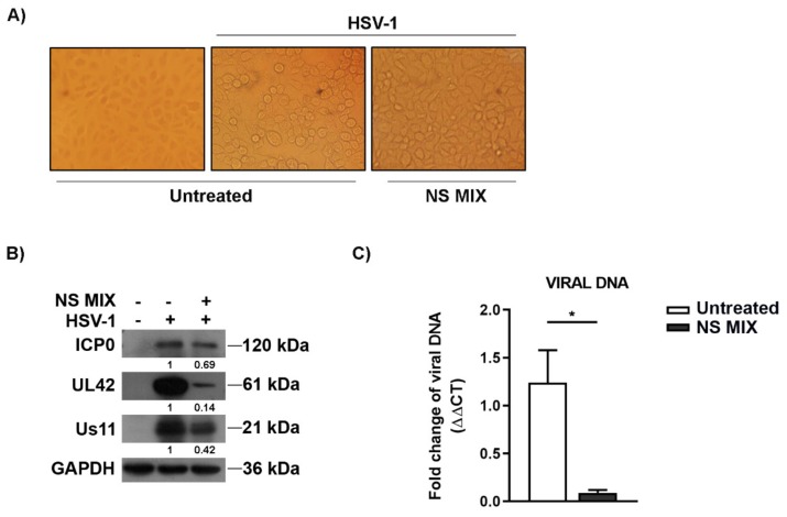
Figure 3.
NS MIX affected the expression of viral antigens and HSV-1 replication. (A) Normal phase contrast inverted micrographs of Vero cells treated with 0.4 mg/mL of NS MIX. The cells were pre-treated with NS MIX for 1 h at 37 °C, then cells either mock infected or infected with HSV-1 at multiplicity of infection (MOI) of 1 for 1 h and incubated in presence of 0.4 mg/mL of the NS MIX for 24 h. (B) Immunoblot analysis was performed to detect α (ICP0), β (UL42), and γ (US11) viral proteins. GAPDH protein was used as loading control. Band density indicated in the figure was determined with the TINA program (version 2.10, Raytest, Straubenhardt, Germany) and expressed as the fold change over the housekeeping gene GAPDH. (C) Relative quantization of viral DNA was performed using real-time quantitative PCR and analyzed by the comparative Ct method (ΔΔCt). Data are expressed as a mean (± SD) of at least three experiments and asterisk (*) indicate the significance of p-values less than 0.05.

