Figure 3.
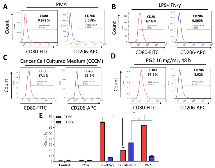
The enhancement of M1 macrophage polarization by PG2 is akin to the effect of LPS/IFN-γ stimulation of MDMs. Images from flow cytometric analyses showing (A) the differentiation of THP-1 monocyte into macrophages after 24 h incubation in PMA; (B) MDMs after exposure to IFN-γ and LPS induced a CD80highCD206low M1 phenotype; (C) cancer cell culture medium (CCCM) induced 17.1% CD80+ and 61.9% CD206+ MDMs; (D) PG2-treatment of MDMs pre-incubated in CCCM induced a CD80highCD206low M1 phenotype, similar to IFN-γ/LPS exposure; (E) a graphical representation of the differential effect of IFN-γ/LPS, CCCM, and 16 mg/mL PG2 treatment on M1–M2 polarization. PG2 enhanced the M1 phenotype akin to IFN-γ/LPS exposure effect. **p < 0.01

