Fig. 5.
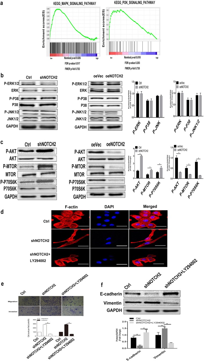
Loss of NOTCH2 promotes EMT in NPC cells by activating AKT signaling. a A GSEA plot showed that the MAPK signaling pathway (P = 0.026 < 0.05) and PI3K signaling pathway (P = 0.010 < 0.05) were gradually inhibited as NOTCH2 expression increased in patients with NPC. b Western blots showing that abnormal expression of NOTCH2 did not alter MAPK signaling component activity. P > 0.05, Student’s t-test. c Western blots showing that NOTCH2 knockdown increased AKT/MTOR/P70s6K signaling component activity and that overexpression of NOTCH2 had the opposite effects. *P < 0.05, Student’s t-test. d Representative images of the cytoskeleton (red) and DAPI (blue) in the indicated CNE-2 cells after 24 h of treatment with the AKT inhibitor LY294002. e Transwell invasion assays showing that the AKT inhibitor reduced shNOTCH2 cell invasion in vitro. *P < 0.05, one-way ANOVA. (f) Compared with the shNOTCH2 group, the shNOTCH2 + LY294002 group had downregulated vimentin expression and upregulated E-cadherin expression. *P < 0.05, one-way ANOVA. Scale bars = 50 μm

