. 2019 Jul 8;10(34):7946–7951. doi: 10.1039/c9sc01410a

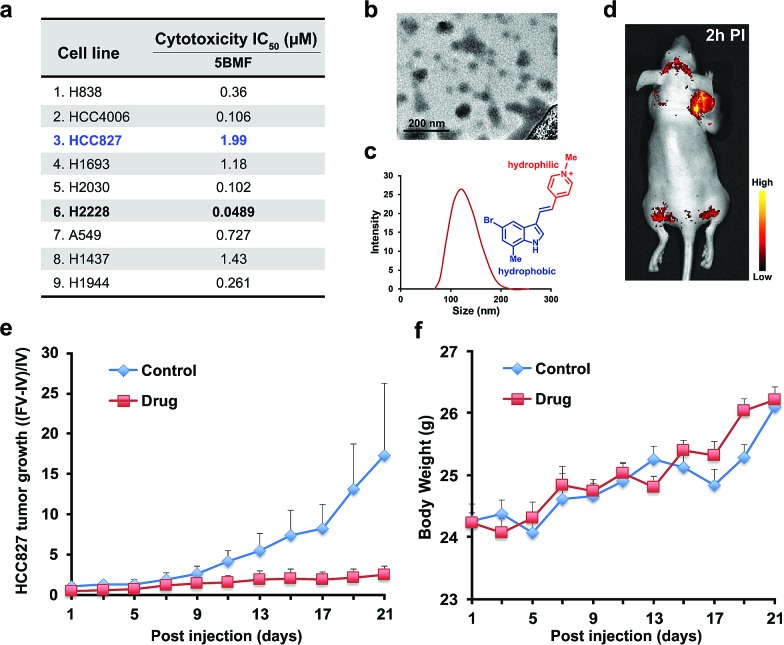
Fig. 5. (a) 5BMF's IC50 of 9 lung cancer cell lines. (b) TEM image of 5BMF, scale bar: 200 nm. (c) DLS curve of 5BMF in PBS buffer, inset image: chemical structure of the amphiphilic 5BMF with the hydrophilic N-methylpyridinium group (red) and hydrophobic indole group (blue). (d) A representative in vivo fluorescence image of a human lung cancer HCC827 bearing mouse 2 h post-injection (PI) of 5BMF (15 mg kg–1, 0.306 M kg–1; for a 20 g mouse, inject 6.11 mM 5BMF PBS solution 150 μL; fluorescence imaging was performed with 450 nm excitation light, and the emission light was collected using a 550 nm long-pass filter) (n = 4). (e) Effects of 5BMF on the tumor growth of HCC827 in nude mice (n = 10 per group). FV, final volume. IV, initial volume. (f) Effects of 5BMF on the tumor-bearing nude mice's body weight (n = 10 per group). The treatment was started when the tumors reached ∼3 mm in diameter (day 1). Control group: PBS. Drug group: 5BMF, 15 mg kg–1, 0.306 M kg–1; for a 20 g mouse, inject 6.11 mM 5BMF PBS solution 150 μL; IV injections were given on day 1, 3, 5, 7, 9, 11, 13, 15, 17, 19, and 21.

Fig. 5