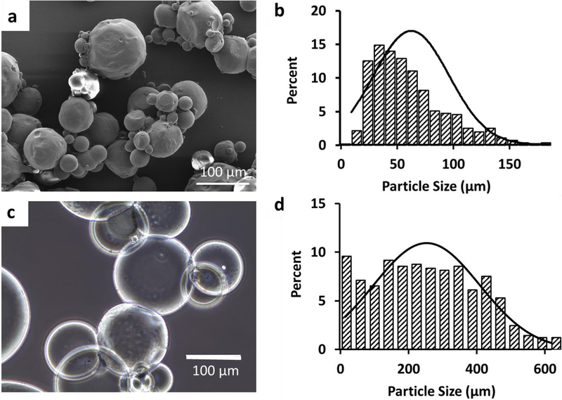
Figure 2:
Gelatin microgels. a) SEM image of lyophilized (dry) microgels. b) Size distribution of the dry microgels. The average diameter of the dry microgels was 63μm (± 34μm). c) Optical microscope image of the gelatin microgels after swelling in PBS. d) Size distribution of the microgels after swelling. The average diameter was 253μm (± 155μm).

