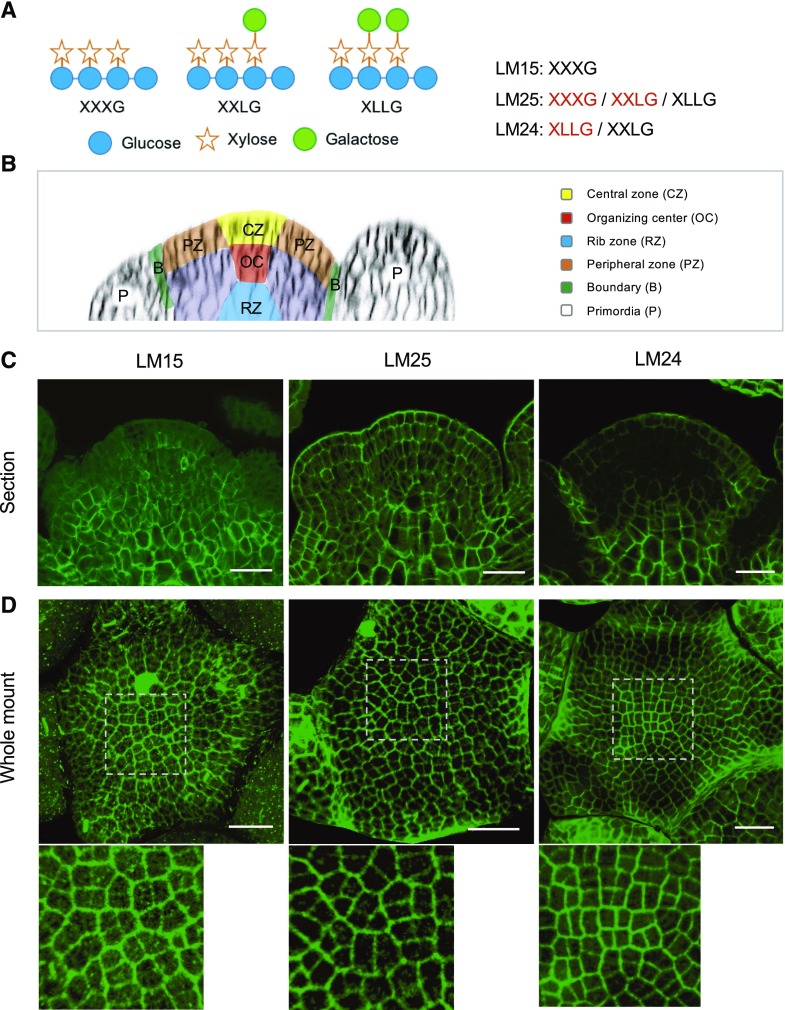
Figure 1.
Differential distribution of xyloglucans (XyGs) in Arabidopsis wild-type shoot apices. A, Schematic structures of XyG subunits and specificity of XyG antibodies. Letters highlighted by red color mean higher affinity. B, Schematic structure of Arabidopsis SAM. C and D, Immunolocalization of XyGs in wild-type (Col) shoot apex sections (C) and whole mount tissues (D) labeled with LM15, LM25, and LM24 antibodies. Details are shown at bottom of (D). Scale bars = 20 µm.

