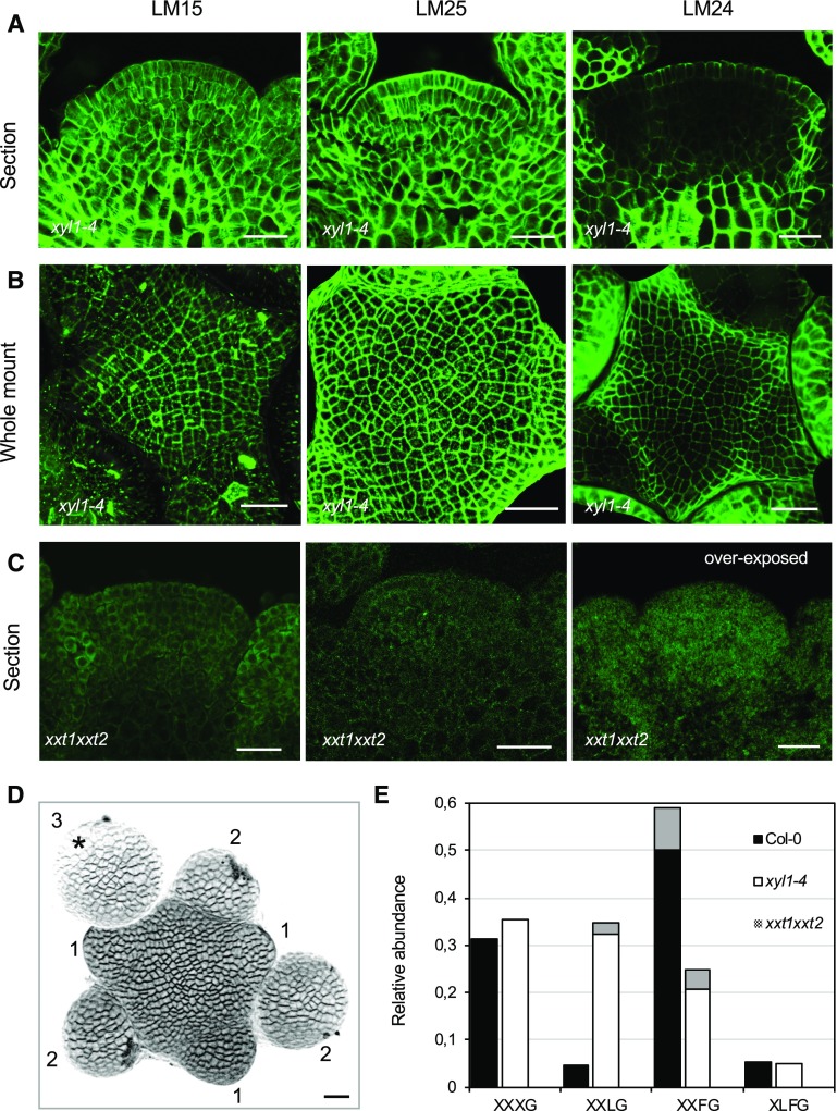
Figure 2.
Altered distribution of XyGs in Arabidopsis XyG mutant shoot apices. A to C, Immunolocalization of XyGs in mutant backgrounds using LM15, LM25, and LM24 antibodies. Sections of xyl1-4 shoot apices (A), whole mount labeling of xyl1-4 (B), and sections of xxt1xxt2 shoot apices (C) are shown. Scale bars = 20 µm. D, Three-dimensional reconstruction image of the shoot apices prepared for XyG composition analysis. The buds are numbered according to their developmental stages (Smyth et al., 1990). Asterisk marks the flower bud at stage 3, which was not included for the sampling. Scale bar = 20 µm. E, MALDI-TOF MS analysis of XyGs in wild-type, xyl1-4, and xxt1xxt2 shoot apices. Gray areas of columns represent the proportion of acetylated subunits.

