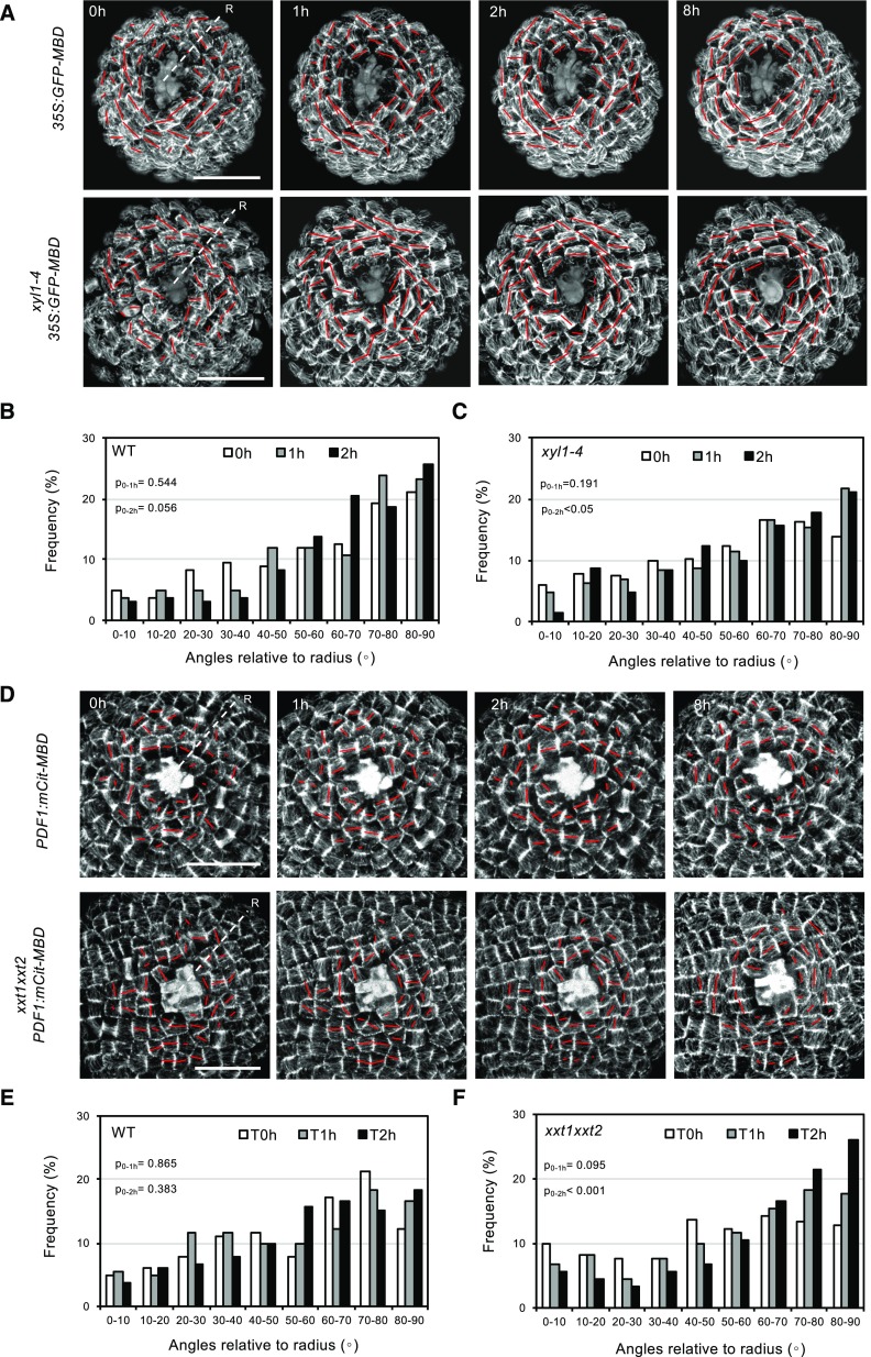
Figure 8.
CMT reactions to mechanical perturbation in wild-type, xyl1-4, and xxt1xxt2 SAMs. A, Time series of CMT patterning in 35S:GFP-MBD (wild type [WT]) and xyl1-4 35S:GFP-MBD SAMs after laser ablation at the meristem center. The orientation and the length of the red bar represent average CMT orientation and degree of CMT anisotropy respectively at cellular level. B and C, Quantification of CMT orientation angles relative to radius of wild type (B) and xyl1-4 (C) SAMs, 1 and 2 h after laser ablation; n = 167 cells from 4 wild-type meristems and n = 204 cells from 4 xyl1-4 meristems. D, Time series of CMT patterning on pPDF1:mCitrine-MBD (wild type) and xxt1xxt2 pPDF1:mCitrine-MBD SAMs after laser ablation at the meristem center. E and F, Quantification of CMT orientation angles relative to the SAM radius of wild type (E) and xxt1xxt2 (F) SAMs, 1 and 2 h after laser ablation; n = 164 cells from 4 wild-type meristems and n = 181 cells from 4 xxt1xxt2 meristems. ‘R’ in (A and D) represents radius of meristem. P-values are calculated based on Kolmogorov-Smirnov test. Scale bars = 20 µm.

