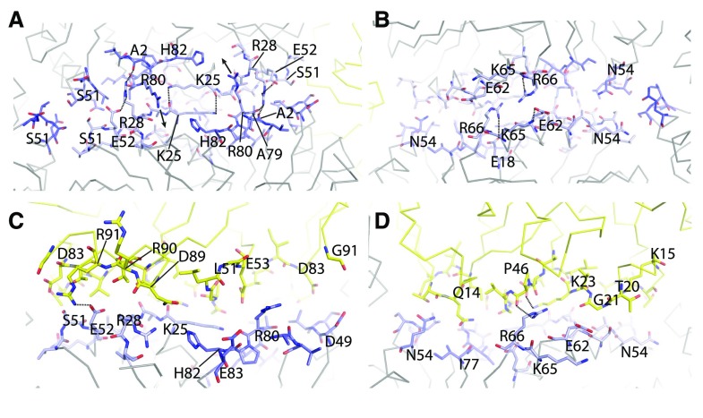
Figure 3.
Protein subunit interfaces. A and B, View onto the CcmK-CcmK interface from the outside (A) and from the inside (B). C and D, View of the CcmK-CcmL interface from the inside (C) and outside (D). Interface residues are shown as sticks, CcmK in shades of blue and CcmL in yellow. Important residues are labeled, and dashed lines indicate specific interactions.

