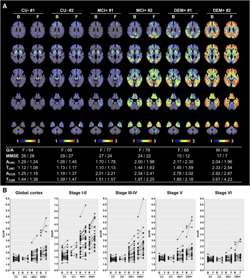
FIGURE 1.
Examples of 18F-flortaucipir PET SUVR images created with cerebellar crus as reference region and their longitudinal changes in PVE-corrected SUVR obtained at baseline and follow-up. (A) Examples of spatially normalized PET images uncorrected for PVE, with 18F-florbetaben and 18F-flortaucipir SUVR and MMSE scores presented below the images. (B) 18F-flortaucipir SUVR measured in global cortical gray matter and composite regions corresponding to different Braak stages. B = baseline; F = follow-up; G/A = sex/age; A and T = global cortical SUVR of 18F-florbetaben or 18F-flortaucipir PET, respectively; COR and UNC = SUVR with and without PVE correction, respectively.

